Plantar Plate Injury: Assessment and Management
By Mr Hussain
Disclaimer: This article is for educational purposes only and is not intended to replace individual medical assessment or advice. If you are experiencing foot pain or injury, consult a qualified healthcare professional.
Introduction
Plantar plate injuries are frequently missed and remain under-diagnosed, particularly in the UK. One key reason is limited awareness of the plantar plate itself. As the saying goes: if you’re not looking for it, you won’t find it. These injuries are often mislabelled as metatarsalgia, a term that merely describes pain in the forefoot rather than identifying its underlying cause.
This article outlines the anatomy, biomechanics, clinical presentation, assessment, imaging, and evidence-based management of plantar plate injuries, drawing on current literature and clinical practice.
What Is the Plantar Plate?
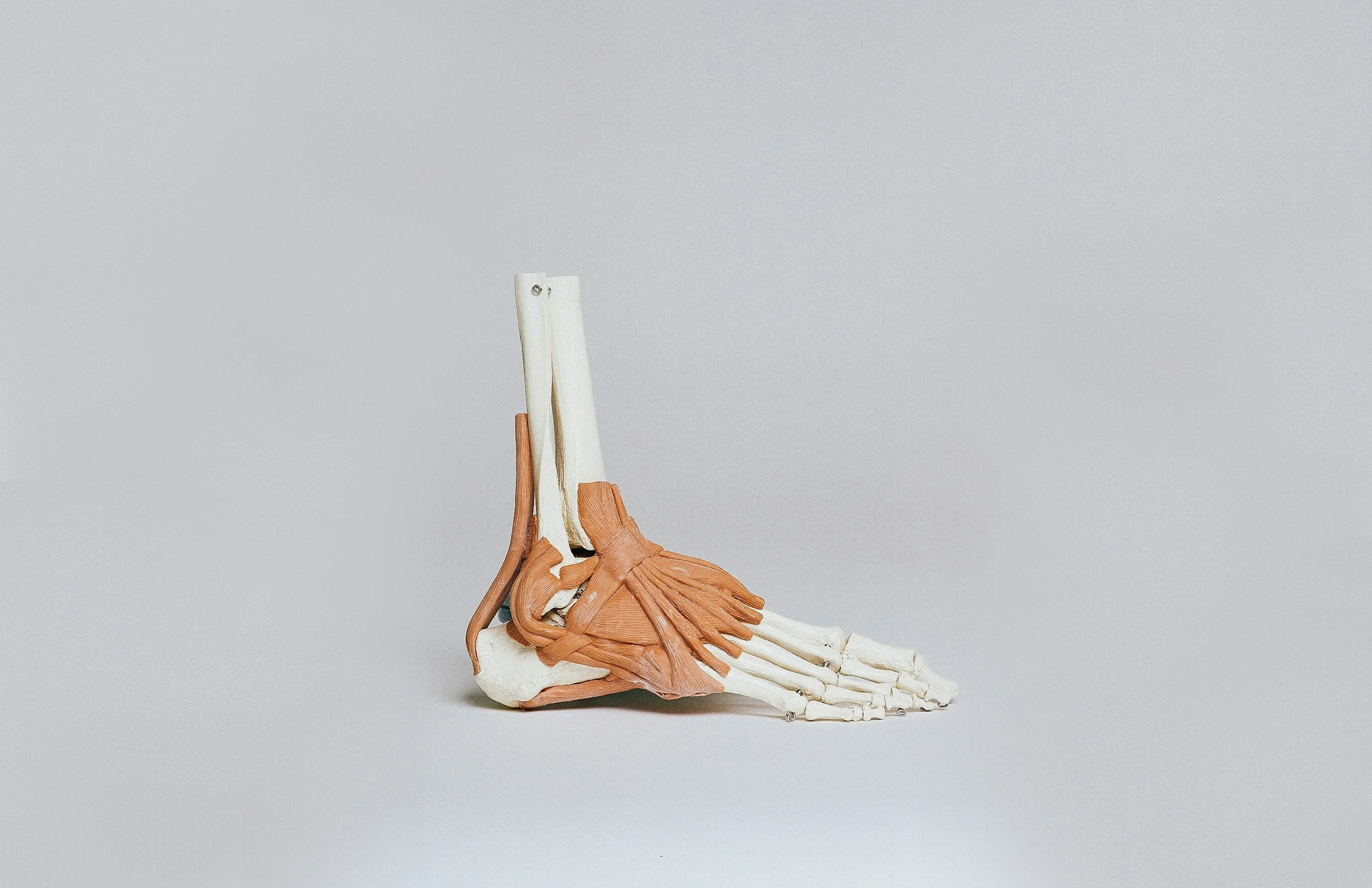
The plantar plate is a fibrocartilaginous structure that originates from the plantar aspect of the metatarsal head and inserts into the base of the proximal phalanx via the joint capsule. Alongside surrounding ligaments and musculature, it plays a critical role in stabilising the metatarsophalangeal joints (MTPJs).
Importantly, the plantar plate also serves as an attachment site for the plantar fascia. During weight-bearing, elongation of the medial arch tensions the plantar fascia, engaging the plantar plate and assisting plantarflexion of the proximal phalanx until toe-off. This interaction is commonly described as the reverse windlass mechanism.¹²
Clinical Assessment: Reverse Windlass Function
A simple and effective way to assess reverse windlass function is the Footstool Edge Test, described by Stainsby.⁴
The patient stands with the MTPJs positioned at the edge of a stool, allowing the toes to hang freely. In a functioning reverse windlass, the proximal phalanges plantarflex. Failure to do so may indicate plantar plate dysfunction or rupture.
Causes and Prevalence
Plantar plate injury is one of the most common causes of second MTPJ pain.⁵ Improved imaging techniques have led to increased detection rates.⁶⁷
Contributing factors include:
Repetitive or excessive MTPJ dorsiflexion (e.g. running, jumping, dancing)
Forefoot-dominant running styles
Hallux valgus, leading to reduced first MTPJ function and compensatory loading of lesser toes
Metatarsal length abnormalities
Footwear factors such as high heels or flexible minimalist shoes
Anything that increases ground reaction force or dorsiflexion stress at the MTPJs may overload the plantar plate.
Clinical Presentation
Patients typically report:
Dorsal and plantar MTPJ pain described as aching or bruising
Pain aggravated by weight-bearing (running, dancing, barefoot walking)
Symptom relief with rest or non-weight-bearing
Worsening symptoms in flexible shoes or high heels
Reduced plantarflexion strength (Digital Purchase Test)
Localised oedema
Positive Digital Lachman’s / Vertical Stress Test
Late-stage signs such as floating toe, hammertoe, or the Churchill sign
Digital Lachman’s (Vertical Stress) Test
This test assesses plantar plate integrity in a similar manner to ACL testing at the knee. The metatarsal head is stabilised while a vertical force is applied to the proximal phalanx.
Sensitivity: 80.6%
Specificity: 99.8%⁸
Pain and excessive dorsal translocation suggest plantar plate pathology.
Classification Systems
Two commonly used grading systems exist:
Thompson & Hamilton⁹
Stage 0: No dorsal translocation
Stage 1: Subluxation without dislocation
Stage 2: Reducible dislocation
Stage 3: Fixed dislocation
Yu & Judge¹⁰
Stage 1: Plantar tenderness, no deformity
Stage 2: Moderate oedema with deviation
Stage 3: Persistent deformity with subluxation or dislocation
In clinical practice, Thompson & Hamilton better reflect structural integrity, while Yu & Judge focus on observable clinical progression.
Imaging
There is ongoing debate regarding optimal imaging modality:
MRI: Sensitivity 95%, specificity 54%
Ultrasound: Sensitivity 93%, specificity 33%¹¹
MRI provides broader structural context, while ultrasound is more operator-dependent but cost-effective.
Weight-bearing X-rays can reveal dorsal subluxation and help exclude bony pathology.¹²
Notably, asymptomatic plantar plate tears are common—one study identified injuries in 35% of asymptomatic individuals.¹³ This reinforces the importance of treating the patient, not the scan.
Management Principles
The primary goal of treatment is load management:
Reduce ground reaction force beneath the affected metatarsal
Limit excessive dorsiflexion of the proximal phalanx
Conservative Treatment Options
Activity modification: Avoid barefoot walking
Footwear: Stiff-soled shoes, reduced heel height
Strapping: Digit plantarflexion taping using rigid zinc oxide tape
Orthoses:
Metatarsal dome proximal to the affected head
Sulcus extension with MTPJ cut-out
Strength & flexibility:
Calf flexibility to reduce forefoot overload
Intrinsic foot muscle strengthening
Injection Therapy
Corticosteroid injections may be considered cautiously:
Maximum of 3 injections per year
At intervals of 1–3 months
Repeated injections have been associated with:
Reduced tensile strength of ligamentous tissue¹⁶
Increased risk of MTPJ dislocation¹²¹⁴¹⁵
Mr Hussain’s Clinical Approach
No barefoot walking for at least 6 weeks
Stiff-soled footwear
Digit strapping (changed every 72 hours)
Activity modification
Custom orthoses
Progressive strength and mobility work
Regular review and surgical referral only if conservative care fails
Conclusion
Plantar plate injury is a complex but manageable condition when recognised early. Understanding the biomechanics and applying structured conservative management may reduce progression to deformity and the need for surgery.
References
Hicks JH. The foot as a support. Acta Anat. 1955;25(1):34–45
🔗 https://pubmed.ncbi.nlm.nih.gov/13301184/Hicks JH. The mechanics of the foot. II. The plantar aponeurosis and the arch. J Anat. 1954;88:25–30
🔗 https://www.scirp.org/reference/referencespapers?referenceid=2802682Kirby KA. Foot and Lower Extremity Biomechanics III: Precision Intricast Newsletters, 2002–2008. Precision Intricast, Inc.; 2009
Stainsby GD. Pathological anatomy and dynamic effect of the displaced plantar plate… Ann R Coll Surg Engl. 1997;79(1):58–68
🔗 https://pmc.ncbi.nlm.nih.gov/articles/PMC2502622/Yao L, Do HM, Cracchiolo A, Farahani K. Plantar plate of the foot… AJR Am J Roentgenol. 1994;163(3):641–644
Ashman CJ, Klecker RJ, Yu JS. Forefoot pain involving the metatarsal region… Radiographics. 2001;21(6):1425–1440
Coughlin MJ. Second metatarsophalangeal joint instability in the athlete. Foot Ankle. 1993;14(6):309–319
🔗 https://journals.sagepub.com/doi/abs/10.1177/107110079301400601Klein EE, Weil L, Weil LS, Coughlin MJ. Clinical Examination of Plantar Plate Abnormality… Foot Ankle Int. 2013; DOI:10.1177/1071100712471825
🔗 https://pubmed.ncbi.nlm.nih.gov/?term=1071100712471825Thompson FM, Hamilton WG. Problems of the second metatarsophalangeal joint. Orthopedics. 1987;10(1):83–89
🔗 https://pubmed.ncbi.nlm.nih.gov/3809019/ PubMedYu GV, Judge MA. Predislocation Syndrome of the Lesser Metatarsophalangeal Joint… In: Reconstructive Surgery of the Foot and Leg – Update ’95
Duan X, Li L, Wei D-Q, et al. Role of MRI versus ultrasound for detection of plantar plate tear. J Orthop Surg Res. 2017;12(1):14
🔗 https://pubmed.ncbi.nlm.nih.gov/28109309/Yu GV, Judge MS, Hudson JR, Seidelmann FE. Predislocation syndrome… J Am Podiatr Med Assoc. 2002;92(4):182–199
Gregg J, Silberstein M, Schneider T, Marks P. Sonographic and MRI evaluation of the plantar plate… Eur Radiol. 2006
Reis ND, Karkabi S, Zinman C. Metatarsophalangeal joint dislocation after steroid injection. J Bone Joint Surg Br. 1989;71(5):864
🔗 https://www.koreamed.org/SearchBasic.php?RID=2539689Gray RG, Gottlieb NL. Intra-articular steroids: an updated assessment. Clin Orthop. 1983;177:235
Noyes FR, Nussbaum NS, Torvik PJ, Cooper S. Changes in ligaments after corticosteroid injections. J Bone Joint Surg. 1975;57:876